Leica SP8
Room B:10, BMIC, Synergy LG.
The Leica SP8 is a high-performance upright microscope capable of the following:
-
Laser-Scanning Confocal imaging, to image engineered- and auto-fluorescence with high resolution and contrast.
-
Multiphoton imaging (2-Photon excitation microscopy), to image thicker tissues (up to 1mm).
-
Fluorescence-Lifetime Imaging Microscopy (FLIM), to image protein-protein interactions or specific compounds.
-
Second-Harmonic imaging microscopy, to image crystaline structures within tissues.
Laser lines available
Imaging mode |
Name |
Excitation Wavelength |
Confocal |
UV |
405nm |
Argon |
458nm |
|
476nm |
||
488nm |
||
496nm |
||
514nm |
||
White Light Laser (WLL) |
470-670nm (tunable in 1nm increments) |
|
Multiphoton |
Insight DeepSee #1 line |
1040nm |
Insight DeepSee #2 line |
680-1300nm (tunable) |
Detectors available
Name |
Number available |
Advantage |
Photomultiplier Tube (PMT) |
3 |
Very robust – always start with these. |
Hybrid Detector (HyD) |
2 |
Very sensitive to damage by strong signal but ideal for capturing dim signal – only use when all else fails, to avoid damage. |
Scanners available
Name |
Advantage |
Default Scanner |
Very versatile – the scanner used 99% of the time. |
Resonant Scanner |
Limited control of position and speed control but very fast, so ideal for imaging very fast moving organelles in living tissue. |
The system is controlled using LASX software platform and allowing multiple post-processing options including 3D modelling and Huygens deconvolution software.
Training: use of this microscope requires at least two training sessions with Viv or Phil. Please contact us at least 2 weeks in advance to organise training. Registration to book the equipment will not be available until training is complete.
Image gallery – example images from the Leica SP8
Madeline Mitchell and Vivien Rolland - Laser-scanning confocal micrograph of a cotton leaf. Have we made crease-free cotton? After three years, the CSIRO cotton biotech team celebrated the successful engineering of a new stretchy building block into cotton cell walls (pink). We hope this will enhance cotton fibre properties so that one day our shirts won’t need ironing! Multichannel confocal imaging of a cotton leaf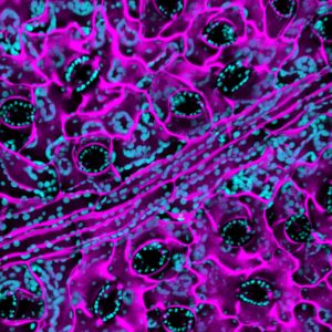
Vivien Rolland Photography competition Multichannel confocal imaging of a tobacco leaf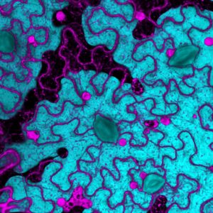
Phil Hands - 3D reconstruction of a series of laser-scanning confocal micrographs of L. maculans fungal hyphae surrounding the vasculature inside a Canola cotyledon. 3D reconstruction of fungal hyphae in a canola leaf
Rosemary White - Second harmonic imaging of cotton fibres. 2nd harmonic imaging of cotton fibres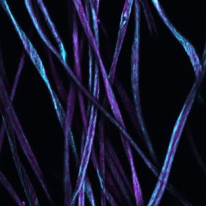
A BMIC user manual is available via the EZbooking object page
Additional resource downloads
SP8 operation thesis – Download